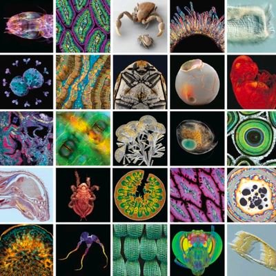
Search results for #Micrograph

Congrats to our grad student Jade Fisher, finalist in #ScienceExposed 2025! 📸 Her research explores how a rare genetic disorder causes neurons to break down over time. 🗳️ Vote for her #micrograph today & help Jade bring home a #URegina win! @NSERC_CRSNG @FGSR_UofR #GoFarUofR

Geometric elegance in a grain of dandelion pollen #SEM #micrograph #sphere


Rotifer Polyarthra sp. 150 µm long showing heart-shaped ovary with nuclei and yolk. Focus stacking with light microscopy in differential interference contrast (DIC). Submitted by: Håkan Kvarnström, , Bromma, Sweden #MicroscopyMonday #Micrograph

Mold (likely Aspergillus sp.) on a plum seed. Focus stack with reflected light micrograph. 🔬 Submitted by: Sergii Dymchenko, SDym Photography, Bellevue, WA #MicroscopyMonday #Micrograph


🦠This scanning electron #micrograph shows Gram-negative "Flexispira rappini" #bacteria, magnified 13,951x. The name is provisional, and it is now referred to as #Helicobacter sp. #flexispira in the literature due to its relation to #Helicobacter spp.🔗encyclopedia.pub/image/detail/1…


Image of the Week: Mayfly Beauty Micrograph of tracheae and tracheoles in the gills of a mayfly larva, taken in polarized light and dark field. Read more here: ow.ly/JRFG50yThjQ #Micrograph #tracheae #tracheoles #mayfly #larva #polarized #light


The #framework #structure of AlPO-18 (a), SEM #micrograph of AlPO-18 (b), #Nitrogen #adsorption/#desorption isotherm (c), and temperature-dependent in situ powder X-ray #diffraction of (as-synthesized) AlPO-18 (d)

Calling all #microscopists! The submission deadline (March 15th) is quickly approaching for MT's 5th annual #Micrograph Competition. We are excited to see all the beautiful images you submit this year! #microscopy microscopy.org/microscopy-tod…


A close up #micrograph of the #infestations on your skin. 🔬😱 #AI #AIArt #AIArtCommunity #AIArtist #AIArtistCommunity #AIArtwork #ComputerArt #DigitalArt

Originally each pixel in this #micrograph was tied to the signal gathered as the beam traveled over a position on the sample.

'Fog City'🏙️Selective area grown InP nano-pillars covered in photoresist! 2nd prize winner of the 2020 NEMI photo competition!🥈 📸Imaged by Jason Jung, @TUeindhoven 🔬#SEM #micrograph on JEOL JSM-7500FA, Eindhoven (@TUeindhoven) @ProjectNemi #Microscopy #ElectronMicroscopy


This TEM #micrograph shows strange structure inside a #cell! #Microscopy #ElectronMicroscope

Happy #MicroscopyMonday! 'Fog City'🏙️showing selective area grown InP semiconductor pillars covered in photoresist!📸Jason Jung 🔬#SEM #micrograph on JEOL JSM-7500FA at TU Eindhoven @TUeindhoven @ProjectNemi


(´-ω-`) @micrographMid
1 Followers 0 Following 以前アメリカにいた時は写真家。今は電子書籍のプロデューサーや、集客のコンサルとかやってます。 『人生一度きり』をモットーに、自由に働き、自分らしい人生をおくりたい人を支援するような活動をしていきたいと思っています。どなたでもフォロー大歓迎です^^♪
n2d7w7a j4s1z5 @Micrograph582
1 Followers 0 Following
micrograph tfh @Micrograph50841
1 Followers 0 Following
c1v3v9c w1n4n5u @Micrograph63
1 Followers 0 Following
micrograph 每日大�... @Micrograph82053
1 Followers 1 Following
micrograph 每日大�... @Micrograph29476
1 Followers 1 Following
Sam's Micrographs @Micrograph66836
2 Followers 4 Following
Micrograph @micrograph_it
11 Followers 35 Following
i2p3p3s h3x3p6 @Micrograph477
1 Followers 8 Following